
Search
- Page Path
- HOME > Search
Original Articles
- Diabetes, obesity and metabolism
- Coronary Artery Calcium Score as a Sensitive Indicator of Cardiovascular Disease in Patients with Type 2 Diabetes Mellitus: A Long-Term Cohort Study
- Dae-Jeong Koo, Mi Yeon Lee, Sun Joon Moon, Hyemi Kwon, Sang Min Lee, Se Eun Park, Cheol-Young Park, Won-Young Lee, Ki Won Oh, Sung Rae Cho, Young-Hoon Jeong, Eun-Jung Rhee
- Endocrinol Metab. 2023;38(5):568-577. Published online October 10, 2023
- DOI: https://doi.org/10.3803/EnM.2023.1770

- 1,605 View
- 114 Download
-
 Abstract
 PDF
PDF Supplementary Material
 PubReader
  ePub
ePub - Background
Coronary artery calcium score (CACS) has become an important tool for evaluating cardiovascular disease (CVD). This study evaluated the significance of CACS for future CVD through more than 10 years of follow-up in asymptomatic Korean populations with type 2 diabetes mellitus (T2DM) known to have a relatively low CACS burden.
Methods
We enrolled 981 asymptomatic T2DM patients without CVD at baseline who underwent CACS evaluation using multidetector computed tomography between January 2008 and December 2014. They were grouped into five predefined CACS categories based on Agatston scores and followed up by August 2020. The primary endpoint was incident CVD events, including coronary, cerebrovascular, and peripheral arterial disease.
Results
The relative risk of CVD was significantly higher in patients with CACS ≥10, and the significance persisted after adjustment for known confounders. A higher CACS category indicated a higher incidence of future CVD: hazard ratio (95% confidence interval) 4.09 (1.79 to 9.36), 12.00 (5.61 to 25.69), and 38.79 (16.43 to 91.59) for 10≤ CACS <100, 100≤ CACS <400, and CACS ≥400, respectively. During the 12-year follow-up period, the difference in event-free survival more than doubled as the category increased. Patients with CACS below 10 had very low CVD incidence throughout the follow-up. The receiver operating characteristic analysis showed better area under curve when the CACS cutoff was 10 than 100.
Conclusion
CACS can be a sensitive marker of CVD risk. Specifically, CACS above 10 is an indicator of CVD high-risk requiring more intensive medical treatment in Koreans with T2DM.

- Clinical Study
- Serum Transferrin Predicts New-Onset Type 2 Diabetes in Koreans: A 4-Year Retrospective Longitudinal Study
- Jong Dai Kim, Dong-Mee Lim, Keun-Young Park, Se Eun Park, Eun Jung Rhee, Cheol-Young Park, Won-Young Lee, Ki Won Oh
- Endocrinol Metab. 2020;35(3):610-617. Published online September 22, 2020
- DOI: https://doi.org/10.3803/EnM.2020.721
- 4,412 View
- 98 Download
- 5 Web of Science
- 5 Crossref
-
Abstract
PDFPubReader ePub
- Background
It is well known that high serum ferritin, a marker of iron storage, predicts incident type 2 diabetes. Limited information is available on the association between transferrin, another marker of iron metabolism, and type 2 diabetes. Thus, we investigated the association between transferrin and incident type 2 diabetes.
Methods
Total 31,717 participants (mean age, 40.4±7.2 years) in a health screening program in 2005 were assessed via cross-sectional analysis. We included 30,699 subjects who underwent medical check-up in 2005 and 2009 and did not have type 2 diabetes at baseline in this retrospective longitudinal analysis.
Results
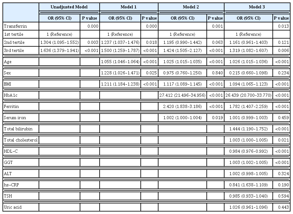
The serum transferrin level was higher in the type 2 diabetes group than in the non-type 2 diabetes group (58.32±7.74 μmol/L vs. 56.17±7.96 μmol/L, P<0.001). Transferrin correlated with fasting serum glucose and glycosylated hemoglobin in the correlational analysis (r=0.062, P<0.001 and r=0.077, P<0.001, respectively) after full adjustment for covariates. Transferrin was more closely related to homeostasis model assessment of insulin resistance than to homeostasis model assessment of β cell function (r=0.042, P<0.001 and r=–0.019, P=0.004, respectively) after full adjustment. Transferrin predicted incident type 2 diabetes in non-type 2 diabetic subjects in a multivariate linear regression analysis; the odds ratio (95% confidence interval [CI]) of the 3rd tertile compared to that in the 1st tertile of transferrin for incident diabetes was 1.319 (95% CI, 1.082 to 1.607) after full adjustment (P=0.006).
Conclusion
Transferrin is positively associated with incident type 2 diabetes in Koreans. -
Citations
Citations to this article as recorded by
- Plasma proteome profiling reveals the therapeutic effects of the PPAR pan-agonist chiglitazar on insulin sensitivity, lipid metabolism, and inflammation in type 2 diabetes
Xingyue Wang, You Wang, Junjie Hou, Hongyang Liu, Rong Zeng, Xiangyu Li, Mei Han, Qingrun Li, Linong Ji, Desi Pan, Weiping Jia, Wen Zhong, Tao Xu
Scientific Reports.2024;[Epub] CrossRef - Plasma Proteomic Signature of Endometrial Cancer in Patients with Diabetes
Muhammad Mujammami, Mohamed Rafiullah, Khalid Akkour, Assim A. Alfadda, Afshan Masood, Salini Scaria Joy, Hani Alhalal, Maria Arafah, Eman Alshehri, Ibrahim O. Alanazi, Hicham Benabdelkamel
ACS Omega.2024; 9(4): 4721. CrossRef - Association between systemic iron status and β-cell function and insulin sensitivity in patients with newly diagnosed type 2 diabetes
Yao Qin, Yiting Huang, Yuxiao Li, Lu Qin, Qianying Wei, Xin Chen, Chuanhui Yang, Mei Zhang
Frontiers in Endocrinology.2023;[Epub] CrossRef - Association of Body Iron Metabolism with Type 2 Diabetes Mellitus in Chinese Women of Childbearing Age: Results from the China Adult Chronic Disease and Nutrition Surveillance (2015)
Jie Feng, Xiaoyun Shan, Lijuan Wang, Jiaxi Lu, Yang Cao, Lichen Yang
Nutrients.2023; 15(8): 1935. CrossRef - Serum Level of Ceruloplasmin, Angiotensin-Converting Enzyme and Transferrin as Markers of Severity in SARS-CoV-2 Infection in Patients with Type 2 Diabetes
Patricia-Andrada Reștea, Ștefan Țigan, Laura Grațiela Vicaș, Luminița Fritea, Eleonora Marian, Tunde Jurca, Annamaria Pallag, Iulius Liviu Mureșan, Corina Moisa, Otilia Micle, Mariana Eugenia Mureșan
Microbiology Research.2023; 14(4): 1670. CrossRef
- Plasma proteome profiling reveals the therapeutic effects of the PPAR pan-agonist chiglitazar on insulin sensitivity, lipid metabolism, and inflammation in type 2 diabetes
- Obesity and Metabolism
- Association between Serum Albumin, Insulin Resistance, and Incident Diabetes in Nondiabetic Subjects
- Ji Cheol Bae, Sung Hwan Seo, Kyu Yeon Hur, Jae Hyeon Kim, Myung-Shik Lee, Moon Kyu Lee, Won Young Lee, Eun Jung Rhee, Ki Won Oh
- Endocrinol Metab. 2013;28(1):26-32. Published online March 25, 2013
- DOI: https://doi.org/10.3803/EnM.2013.28.1.26
- 4,629 View
- 42 Download
- 36 Crossref
-
Abstract
PDFPubReader
Background Serum albumin has been suggested to be associated with insulin resistance. We evaluated the association between serum albumin concentration and insulin resistance. We also investigated whether serum albumin level has an independent effect on the development of diabetes.
Methods In our study, 9,029 subjects without diabetes, who underwent comprehensive health check-ups annually for 5 years, were categorized into tertiles based on their serum albumin levels at baseline. The odds ratio (OR) for the prevalence of insulin resistance, defined as the top quartile of homeostasis model assessment of insulin resistance and the presence of impaired fasting glucose and nonalcoholic fatty liver disease, was evaluated cross-sectionally. Also, the hazard ratio (HR) for incident diabetes was estimated longitudinally, according to the baseline albumin tertiles using Cox proportional hazard analysis respectively.
Results From the lowest to the highest tertile of albumin, the multivariable-adjusted ORs of insulin resistance increased significantly in both men and women. During the mean follow-up period of nearly 4 years, 556 (6.1%) subjects progressed to diabetes. The multivariable-adjusted HR (95% confidence interval [CI]) of diabetes in men were 1, 1.09 (95% CI, 0.86 to 1.40), and 1.10 (95% CI, 0.86 to 1.41), respectively, from the lowest to the highest tertiles of baseline albumin. Corresponding values for women were 1, 1.21 (95% CI, 0.66 to 2.21), and 1.06 (95% CI, 0.56 to 2.02), respectively.
Conclusion Our study showed that increased serum albumin level was associated with insulin resistance. However, serum albumin did not have an independent effect on the development of diabetes.
-
Citations
Citations to this article as recorded by- Prevalence of Non-alcoholic Fatty Liver Disease Detected by Computed Tomography in the General Population Compared with Ultrasonography
Yuki Ito, Kentaro Yoshioka, Kazuhiko Hayashi, Yuko Shimizu, Ryo Fujimoto, Ryosuke Yamane, Michiyo Yoshizaki, Go Kajikawa, Taro Mizutani, Hidemi Goto
Internal Medicine.2024; 63(2): 159. CrossRef - Geriatric nutritional risk index is correlated with islet function but not insulin resistance in elderly patients with type 2 diabetes: A retrospective study
Nan Geng, Yaxue Gao, Yuanyuan Ji, Yingchun Niu, Cuijuan Qi, Yunfeng Zhen, Jinhu Chen, Luping Ren
Medicine.2024; 103(11): e37438. CrossRef - Blood Urea Nitrogen to Serum Albumin Ratio as A New Prognostic
Indicator in Critically Ill Patients with Diabetic Ketoacidosis: A Retrospective
Cohort Study
Tingting Hang, Jing Huang, Guiping He, Jin Li, Tingting Tao
Experimental and Clinical Endocrinology & Diabetes.2024;[Epub] CrossRef - Sex difference in the associations among liver function parameters with incident diabetes mellitus in a large Taiwanese population follow-up study
Yi-Kong Chen, Pei-Yu Wu, Jiun-Chi Huang, Szu-Chia Chen, Jer-Ming Chang
Frontiers in Public Health.2023;[Epub] CrossRef - Antidiabetic Properties of Nymphaea Species (Water Lilies): A Review
A. H. M. Safayet Ullah Prodhan, Farzana Sharmin Mridu
The Natural Products Journal.2023;[Epub] CrossRef - Gestational diabetes in women living with HIV in the UK and Ireland: insights from population‐based surveillance data
Laurette L. Bukasa, Mario Cortina‐Borja, Helen Peters, Graham P. Taylor, Claire Thorne
Journal of the International AIDS Society.2023;[Epub] CrossRef - Cancer and Diabetes: Predictive Factors in Patients with Metabolic Syndrome
Mihai Cosmin Stan, Daniel Georgescu, Ciprian Camil Mireștean, Florinel Bădulescu
Diagnostics.2023; 13(16): 2647. CrossRef - Role of liver parameters in diabetes mellitus – a narrative review
Sana Rafaqat, Aqsa Sattar, Amber Khalid, Saira Rafaqat
Endocrine Regulations.2023; 57(1): 200. CrossRef - Differential cellular responses to FDA-approved nanomedicines: an exploration of albumin-based nanocarriers and liposomes in protein corona formation
Athika Darumas Putri, Ming-Jen Hsu, Chia-Li Han, Fang-Ching Chao, Chun-Hua Hsu, Christian D. Lorenz, Chien-Ming Hsieh
Nanoscale.2023; 15(44): 17825. CrossRef - Association of the HALP Score with Dyslipidemia: A Large, Nationwide Retrospective Study
Yazeed Alshuweishi, Ahmed M. Basudan, Mohammed Alfaifi, Hussam Daghistani, Mohammad A. Alfhili
Medicina.2023; 59(11): 2002. CrossRef - Preventive and Ameliorative Effects of Diet Supplemented with Cucurbita maxima Leaf on Hyperglycemia and Hepatotoxicity in STZ-Induced Diabetic Rats
Job Itanyi Onuche, Arowora Kayode Adebisi , Joseph Ikwebe, Michael Sunday Abu
Asian Journal of Biological Sciences.2023; 16(4): 502. CrossRef - Lower Plasma Albumin, Higher White Blood Cell Count and High-Sensitivity C-Reactive Protein are Associated with Femoral Artery Intima-Media Thickness Among Newly Diagnosed Patients with Type 2 Diabetes Mellitus
Nga Phi Thi Nguyen, Thuc Luong Cong, Thi Thanh Hoa Tran, Binh Nhu Do, Son Tien Nguyen, Binh Thanh Vu, Lan Ho Thi Nguyen, Manh Van Ngo, Hoa Trung Dinh, Hoang Duong Huy, Nghia Xuan Vu, Kien Nguyen Trung, Duong Ngoc Vu, Nghia The Pham, Tuan Dinh Le
International Journal of General Medicine.2022; Volume 15: 2715. CrossRef - Liver-function parameters are associated with incident hypertension in a large Taiwanese population follow-up study
Yi-Hsueh Liu, Szu-Chia Chen, Wen-Hsien Lee, Ying-Chih Chen, Jiun-Chi Huang, Pei-Yu Wu, Chih-Hsing Hung, Chao-Hung Kuo, Ho-Ming Su
Journal of Human Hypertension.2022; 37(6): 496. CrossRef - Can probiotic, prebiotic, and synbiotic supplementation modulate the gut-liver axis in type 2 diabetes? A narrative and systematic review of clinical trials
Yousef Al-Najjar, Maryam Arabi, Pradipta Paul, Ali Chaari
Frontiers in Nutrition.2022;[Epub] CrossRef - Albumin infusion ameliorates liver injury in streptozotocin-induced diabetic rats
CS Bae, T Ahn
Veterinární medicína.2022; 67(5): 245. CrossRef - Ameliorative effect of Annona reticulata L. leaf extract on antihyperglycemic activity and its hepato-renal protective potential in streptozotocin induced diabetic rats
Vineela Pulivarthi, Josthna P., C.V. Naidu
Journal of Ayurveda and Integrative Medicine.2021; 12(3): 415. CrossRef - MALDI-TOF MS Characterisation of the Serum Proteomic Profile in Insulin-Resistant Normal-Weight Individuals
Katarzyna Pastusiak, Eliza Matuszewska, Dagmara Pietkiewicz, Jan Matysiak, Pawel Bogdanski
Nutrients.2021; 13(11): 3853. CrossRef - Insulin sensitivity variations in apparently healthy Arab male subjects: correlation with insulin and C peptide
Noor Suleiman, Meis Alkasem, Shaimaa Hassoun, Ibrahem Abdalhakam, Ilham Bettahi, Fayaz Mir, Manjunath Ramanjaneya, Jayakumar Jerobin, Ahmad Iskandarani, Tareq A Samra, Prem Chandra, Monica Skarulis, Abdul Badi Abou-Samra
BMJ Open Diabetes Research & Care.2021; 9(2): e002039. CrossRef - U-shaped association between serum albumin and development of chronic kidney disease in general hypertensive patients
Chongfei Jiang, Binyan Wang, Youbao Li, Liling Xie, Xianglin Zhang, Jiancheng Wang, Yaren Yu, Yun Song, Min Liang, Guobao Wang, Jianping Li, Yan Zhang, Lishun Liu, Chengzhang Liu, Genfu Tang, Yong Huo, Xiping Xu, Xianhui Qin
Clinical Nutrition.2020; 39(1): 258. CrossRef - Serum albumin cysteine trioxidation is a potential oxidative stress biomarker of type 2 diabetes mellitus
Selvam Paramasivan, Sunil S. Adav, SoFong Cam Ngan, Rinkoo Dalan, Melvin Khee-Shing Leow, Hee Hwa Ho, Siu Kwan Sze
Scientific Reports.2020;[Epub] CrossRef - Serum albumin, cardiometabolic and other adverse outcomes: systematic review and meta-analyses of 48 published observational cohort studies involving 1,492,237 participants
Samuel Seidu, Setor K. Kunutsor, Kamlesh Khunti
Scandinavian Cardiovascular Journal.2020; 54(5): 280. CrossRef - Skeletal muscle reprogramming by breast cancer regardless of treatment history or tumor molecular subtype
Hannah E. Wilson, David A. Stanton, Cortney Montgomery, Aniello M. Infante, Matthew Taylor, Hannah Hazard-Jenkins, Elena N. Pugacheva, Emidio E. Pistilli
npj Breast Cancer.2020;[Epub] CrossRef - Prevalence of nutritional deficiencies in bariatric surgery candidates and its effect on metabolic status
Sílvia Cristina de Sousa Paredes, Fernando Mota-Garcia
Hormones.2020; 19(4): 505. CrossRef The Product of Red Blood Cells and Hematocrit Can Be Used as a Novel Indicator of Impaired Fasting Blood Glucose Status
Ling Feng, Haishan Chen, Jianhui Chen, Chongxiang Xiong, Xiaofei Shao, Xin Wang, Jing Ning, Zhicong Xiang, Xuan Wang, Tong Chen, Hua Xiao, Hongjuan Tang, Xiaolin Li, Guobao Hong, Hequn Zou
Diabetes, Metabolic Syndrome and Obesity: Targets and Therapy.2020; Volume 13: 4007. CrossRef- Thiol/Disulphide homeostasis, ischemia modified albumin, and ferroxidase as oxidative stress markers in women with obesity with insulin resistance
Elif Ates, Turan Set, Süleyman Caner Karahan, Cemile Biçer, Özcan Erel
Journal of Medical Biochemistry.2019; 38(4): 445. CrossRef - Insulin resistance and chronic kidney disease progression, cardiovascular events, and death: findings from the chronic renal insufficiency cohort study
Sarah J. Schrauben, Christopher Jepson, Jesse Y. Hsu, F. Perry Wilson, Xiaoming Zhang, James P. Lash, Bruce M. Robinson, Raymond R. Townsend, Jing Chen, Leon Fogelfeld, Patricia Kao, J. Richard Landis, Daniel J. Rader, L. Lee Hamm, Amanda H. Anderson, Har
BMC Nephrology.2019;[Epub] CrossRef - Association of Insulin Based Insulin Resistance with Liver Biomarkers in Type 2 Diabetes mellitus
Usha Adiga, Kathyayani P, Nandith P.B
Journal of Pure and Applied Microbiology.2019; 13(2): 1199. CrossRef - Establishment of an ex Vivo Model of Nonalcoholic Fatty Liver Disease Using a Tissue-Engineered Liver
Qiao Wu, Juan Liu, Lijin Liu, Yu Chen, Jie Wang, Ling Leng, Qunfang Yu, Zhongping Duan, Yunfang Wang
ACS Biomaterials Science & Engineering.2018; 4(8): 3016. CrossRef - Utility of Serum Albumin for Predicting Incident Metabolic Syndrome according to Hyperuricemia
You-Bin Lee, Ji Eun Jun, Seung-Eun Lee, Jiyeon Ahn, Gyuri Kim, Jae Hwan Jee, Ji Cheol Bae, Sang-Man Jin, Jae Hyeon Kim
Diabetes & Metabolism Journal.2018; 42(6): 529. CrossRef - Association of angiotensin-II levels with albuminuria in subjects with normal glucose metabolism, prediabetes, and type 2 diabetes mellitus
Se Hee Min, Sung Hye Kong, Jie-Eun Lee, Dong-Hwa Lee, Tae Jung Oh, Kyoung Min Kim, Kyong Soo Park, Hak Chul Jang, Soo Lim
Journal of Diabetes and its Complications.2017; 31(10): 1499. CrossRef - Three-dimensional perfused human in vitro model of non-alcoholic fatty liver disease
Tomasz Kostrzewski, Terri Cornforth, Sophie A Snow, Larissa Ouro-Gnao, Cliff Rowe, Emma M Large, David J Hughes
World Journal of Gastroenterology.2017; 23(2): 204. CrossRef - Higher serum albumin was related with diabetes incidence and the impact of BMI changes: Based on cohort study of 18,384 Chinese male elderly
Miao Liu, Jingping Tang, Jing Zeng, Yao He
Journal of Diabetes and its Complications.2017; 31(12): 1663. CrossRef - HbA1c as a Screening tool for Ketosis in Patients with Type 2 Diabetes Mellitus
Bing Zhu, Le Bu, Manna Zhang, Aaron M. Gusdon, Liang Zheng, Sharvan Rampersad, Jue Li, Shen Qu
Scientific Reports.2016;[Epub] CrossRef - Association between Serum Albumin Concentration and Ketosis Risk in Hospitalized Individuals with Type 2 Diabetes Mellitus
Po-Chung Cheng, Shang-Ren Hsu, Yun-Chung Cheng
Journal of Diabetes Research.2016; 2016: 1. CrossRef - Brief Review of Articles in 'Endocrinology and Metabolism' in 2013
Won-Young Lee
Endocrinology and Metabolism.2014; 29(3): 251. CrossRef - Serum Albumin Levels: A Simple Answer to a Complex Problem? Are We on the Right Track of Assessing Metabolic Syndrome?
Sohee Kim, Shinae Kang
Endocrinology and Metabolism.2013; 28(1): 17. CrossRef
- Prevalence of Non-alcoholic Fatty Liver Disease Detected by Computed Tomography in the General Population Compared with Ultrasonography
- Isolation of Density Enrichment Fraction of Adipose-Derived Stem Cells from Stromal Vascular Fraction by Gradient Centrifugation Method.
- Min Kyung Kim, Yong Soon Park, Hee Soon Park, Jung Mook Choi, Won Jun Kim, Se Eun Park, Eun Jung Rhee, Cheol Young Park, Won Young Lee, Ki Won Oh, Sung Woo Park, Sun Woo Kim, Kwang Sik Suh, Jeong Taek Woo
- Endocrinol Metab. 2010;25(2):103-109. Published online June 1, 2010
- DOI: https://doi.org/10.3803/EnM.2010.25.2.103
- 2,278 View
- 38 Download
- 1 Crossref
-
Abstract
PDF
- BACKGROUND
Adipose tissues include multipotent cells, the same as bone marrow-derived mesenchymal stem cells. The stromal vascular fractions (SVFs) from adipose tissues represent a heterogeneous cell population. The purpose of this study was to isolate and purify adipose-derived stem cells (ASCs) in SVFs by the density gradient method. METHODS: SVFs were extracted from the subcutaneous, epididymal, mesenteric and retroperitoneal adipose tissue of 8 weeks old male Sprague-Dawley rats (n = 15) and these were separated into 4 layers according to a Nycodenz gradient (Fx-1: < 11%, Fx-2: 11-13%, Fx-3: 13-19% and Fx-4: 19-30%). The post-confluent SVFs were cultured in adipogenic medium for 2 days, in insulin medium for 2 days and in 10% fetal bovine serum medium for 5 days. To observe lipid droplets in SVFs, we performed Oil Red O staining. RESLTS: The SVFs' cellular fractions (Fx-1, Fx-2, Fx-3 and Fx-4) were isolated by density gradient centrifugation from the adipose tissues of rats. The SVFs extracted to fraction 3 (Fx-3) had the most abundant cells compared to that of the other fractions. However fraction 1 (Fx-1) or 2 (Fx-2) had a superior ability to make lipid droplets. The adipogenic differentiation of Fx-1 or 2 was higher than that of the unfractionated cells. The SVFs extracted from retroperitoneal adipose tissue had the highest efficiency for adipogenic differentiation, whereas the SVFs from mesenteric adipose tissue did not differentiate. CONCLUSION: This density gradient fractionated method leads to efficient isolation and purification of cells with the characteristics of ASCs. -
Citations
Citations to this article as recorded by- Embolic Infarction Presented after Intravenous Injection of Stromal Vascular Fraction
Jin Young Seo, Ju-Hun Lee, Hong-Ki Song, Jong Seok Bae, Yerim Kim
Journal of Neurosonology and Neuroimaging.2018; 10(2): 181. CrossRef
- Embolic Infarction Presented after Intravenous Injection of Stromal Vascular Fraction
Retractions of Publication
- Retraction: Relationship between Circulating Osteoprotegerin and Cardiovascular Risk Factors in Women.
- Ki Won Oh, Eun Joo Yun, Eun Sook Oh, Eun Jung Rhee, Won Young Lee, Ki Hyun Baek, Kun Ho Yoon, Moo Il Kang, Cheol Young Park, Moon Ki Choi, Hyung Joon Yoo, Sung Woo Park
- J Korean Endocr Soc. 2008;23(1):69. Published online February 1, 2008
- 1,147 View
- 16 Download
- Retraction: Relationship between Serum Leptin, Adiponectin, Resistin and Ghrelin Levels, and Bone Mineral Density in Men.
- Ki Won Oh, Eun Joo Yun, Eun Jung Rhee, Won Young Lee, Ki Hyun Baek, Kun Ho Yoon, Moo Il Kang, Cheol Young Park, Sung Hee Ihm, Moon Gi Choi, Hyung Joon Yoo, Sung Woo Park
- J Korean Endocr Soc. 2008;23(1):68. Published online February 1, 2008
- 1,053 View
- 16 Download
- Retraction: Relationship between Serum Osteoprotegerin-Receptor Activator of NF-kappaB Ligand Levels and Bone Mineral Metabolism in Men.
- Ki Won Oh, Eun Joo Yun, Eun Jung Rhee, Won Young Lee, Ki Hyun Baek, Moo Il Kang, Cheol Young Park, Sung Hee Ihm, Moon Gi Choi, Hyung Joon Yoo, Sung Woo Park
- J Korean Endocr Soc. 2008;23(1):67. Published online February 1, 2008
- 1,014 View
- 17 Download
Case Report
- A Case of Kallmann's Syndrome Mildly Presenting as Secondary Amenorrhea.
- Na Rae Joo, Cheol Young Park, Hong Ju Moon, Jun Goo Kang, Sung Hee Ihm, Moon Gi Choi, Hyung Joon Yoo, Yul Lee, Ki Won Oh, Sung woo Park
- J Korean Endocr Soc. 2007;22(2):130-134. Published online April 1, 2007
- DOI: https://doi.org/10.3803/jkes.2007.22.2.130
- 2,124 View
- 25 Download
-
Abstract
PDF
- Kallmann's syndrome is very rare congenital defect in GnRH (gonadotrophin releasing hormone) secretion involving both sexes. The mode of inheritance has not been fully understood. But, including X-linked inheritance, the ratio of incidence between male versus female is 5:1, and there is a few case reports of female Kallmann's syndrome in Korea, especially in internal medicine department. We report a case of 35 year-old female Kallmann's syndrome presenting secondary amenorrhea as a mild presentation.
Original Articles
- The Relationship between Lumbar Spine Bone Mineral Density and Cardiovascular Risk Factors in Korean Female Adults.
- Young Yul Koh, Eun Jung Rhee, Se Yeon Kim, Chan Hi Jung, Cheol Young Park, Won Young Lee, Ki Won Oh, Sung Woo Park, Sun Woo Kim
- J Korean Endocr Soc. 2006;21(6):497-505. Published online December 1, 2006
- DOI: https://doi.org/10.3803/jkes.2006.21.6.497
- 2,029 View
- 19 Download
- 4 Crossref
-
Abstract
PDF
- BACKGROUND
Recent studies suggest a possible pathogenic linkage between the osteoporosis and atherosclerosis. We investigated the relationship between cardiovascular risk factors, including high sensitivity C-reactive protein (hs-CRP), insulin resistance, lipid profiles and bone metabolism in Korean females. METHODS: Anthropometric measurements were performed on 437 women (mean age 52 yrs), and cardiovascular risk factors, including fasting blood glucose, fasting blood insulin, lipid profiles and hs-CRP, measured. An atherogenic index was calculated using the serum total cholesterol level divided by the high-density lipoprotein cholesterol (HDL-C) level. The lumbar spine bone mineral density (BMD) was measured using dual X-ray absorptiometry. RESULTS: From bivariate analyses, the lumbar spine BMD showed negative correlations with age, systolic and diastolic blood pressures, serum total cholesterol, low-density lipoprotein cholesterol (LDL-C), triglyceride levels and atherogenic index, and a positive correlation with the HDL-C level. After adjustment for age and BMI, the atherogenic index and HDL-C showed consistent correlation with the lumbar spine BMD. The log-transformed hs-CRP showed no correlation with the lumbar spine BMD. In premenopausal women, age, BMI and atherogenic index showed significant associations with the lumbar spine BMD and the atherogenic index showed consistently significant correlation, even after adjustment for age and BMI. In postmenopausal women, only age and BMI showed significant correlations with the lumbar spine BMD. From multiple linear regression analyses of all the study subjects, age, BMI, atherogenic index and the presence of menopause were found to be determinants of the lumbar spine BMD (R2 = 0.422, p < 0.05), which was consistently significant in analysis performed on premenopausal women (R2 = 0.157, P < 0.05). In postmenopausal women, age and BMI were found to be the determinants of the lumbar spine BMD (R2 = 0.257, P < 0.05). CONCLUSIONS: The lumbar spine BMD was negatively correlated with the atherogenic index in all and in premenopausal women. The menopause seems to play an important role in the relationship of cardiovascular risk factors with BMD in Korean females. -
Citations
Citations to this article as recorded by- Comparison of Relationship between Biochemical Indices and Bone Mineral Densityof Pre- and Post- Menopausal Women in Gyeongnam Area
Mi-Young Park, Sung-Hee Kim
Journal of the East Asian Society of Dietary Life.2017; 27(4): 408. CrossRef - Relationship between Plasma Lipids and Osteoporosis in Korean Postmenopausal Women
Kyung Shik Lee, Jae Hwan Cho, Chang Hae Park, Bo Seung Kim, Kyung Hwan Cho, Seung Hwan Lee, Byung Jun Ko, Do Hoon Kim
Journal of the Korean Geriatrics Society.2011; 15(2): 99. CrossRef - Relationships among Obesity, Bone Mineral Density, and Cardiovascular Risks in Post-menopausal Women
Heeyoung So, Sukhee Ahn, Rhayun Song, Hyunli Kim
Korean Journal of Women Health Nursing.2010; 16(3): 224. CrossRef - Association of the Metabolic Syndrome and Bone Mineral Density in Postmenopausal Women
Jong-Chang Park, Hyuk-Jung Kweon, Yun-Kyo Oh, Hyun-Jin Do, Seung-Won Oh, Youl-Lee Lym, Jae-Kyung Choi, Hee-Kyung Joh, Dong-Yung Cho
Korean Journal of Family Medicine.2010; 31(1): 9. CrossRef
- Comparison of Relationship between Biochemical Indices and Bone Mineral Densityof Pre- and Post- Menopausal Women in Gyeongnam Area
- The Effect of Oxidative Stress on the Proliferation and Differentiation of Human Bone Marrow Stromal Cell-Derived Osteoblasts.
- Eun Sook Oh, Ki Hyun Baek, Won Young Lee, Ki Won Oh, Hye Soo Kim, Je Ho Han, Kwang Woo Lee, Ho Young Son, Sung Koo Kang, Moo Il Kang
- J Korean Endocr Soc. 2006;21(3):222-232. Published online June 1, 2006
- DOI: https://doi.org/10.3803/jkes.2006.21.3.222
- 1,835 View
- 18 Download
- 1 Crossref
-
Abstract
PDF
- BACKGROUND
The objectives of our study were to assess the effects of oxidative stress on the proliferation, differentiation and apoptosis of human bone marrow stromal cell (BMSC)-derived osteoblasts and to explore pathways by which osteoblast cell apoptosis was induced. METHODS: Mononuclear cells including BMSCs were cultured to osteoblastic lineage. Different doses of hydrogen peroxide (H2O2) were added to the culture media. The colony forming units-fibroblastic (CFU-Fs) were stained with crystal violet and alkaline phosphatase (ALP). The MTT assay was done to see the effect of H2O2 on cell viability. The effect of H2O2 on osteocalcin gene expression was determined by RT-PCR. The matrix calcification measurement was performed. FACS analysis was performed to determine the osteoblasts apoptosis. Caspase-3, -8 and 9 activity assay and cytochrome c release were measured. RESULTS: The size and number of ALP (+) CFU-Fs were also decreased by H2O2 treatment. When compared with the control group, H2O2 significantly decreased the total number of cells of each culture well during MTT assay. H2O2 significantly diminished expression of osteocalcin mRNA. N-acetylcystein (NAC) blocked the diminution of cell viability and the inhibition of osteocalcin mRNA expression by H2O2. H2O2 reduced matrix calcification. FACS analysis revealed H2O2 increased percentage of apoptotic cells. Addition of H2O2 resulted in the increase of caspase-9 and -3 activity but not caspase-8, and release of cytochrome c to the cytosol. CONCLUSION: These data suggest that, in primary human BMSCs, oxidative stress inhibits proliferation of stromal cells and inhibits the differentiation to osteoblastic lineage. In addition, oxidative stress induces apoptosis of human BMSC-derived osteoblasts and this may be mediated by mitochondrial pathway of apoptotic signal. -
Citations
Citations to this article as recorded by- Antioxidaitve and Differentiation Effects of Artemisia capillaris T. Extract on Hydrogen Peroxide-induced Oxidative Damage of MC3T3-E1 Osteoblast Cells
Jee-Eun Seo, Eun-Sun Hwang, Gun-Hee Kim
Journal of the Korean Society of Food Science and Nutrition.2011; 40(11): 1532. CrossRef
- Antioxidaitve and Differentiation Effects of Artemisia capillaris T. Extract on Hydrogen Peroxide-induced Oxidative Damage of MC3T3-E1 Osteoblast Cells
- The Relationship of Ghrelin and Leptin with the Biochemical Markers for Adult Growth Hormone Deficiency.
- Chan Hee Jung, Eun Jung Rhee, Se Yeon Kim, Ki Won Oh, Won Young Lee, Sun Woo Kim
- J Korean Endocr Soc. 2006;21(3):213-221. Published online June 1, 2006
- DOI: https://doi.org/10.3803/jkes.2006.21.3.213
- 2,670 View
- 18 Download
-
Abstract
PDF
- BACKGROUND
In spite of the increasing information that has recently been accumulated on the involvement of ghrelin and leptin in energy balance control, the relationship between ghrelin or leptin and the growth hormone (GH)-Insulin like growth factor-1 (IGF-1) axis in the pathological condition characterized by growth hormone deficiency (GHD) has been poorly clarified. Therefore, we performed this study to evaluate the correlation of the plasma levels of ghrelin and leptin with the anthropometric and biochemical markers in GHD adults and also in healthy adults. METHODS: For the 60 male adults (GHD, n = 12; healthy control, n = 48; average age, 54 years), we investigated the correlations between the serum leptin and ghrelin levels with the anthropometric and biochemical factors in the two groups, as divided by their GH status. The diagnosis of GHD was made on the basis of a peak response for serum GH of less than 5 micro/L to a GH provocative test (L-dopa test). All the subjects underwent assessment of waist circumference, BMI and percentage body fat for their body composition. The plasma ghrelin, leptin, insulin, GH and IGF-1 were measured. RESULTS: The groups were well-matched for their age, BMI, waist circumference and percentage of body fat. The ghrelin and leptin levels were not significantly different between the two groups. There was no correlation between the peak GH level or the area under the curve of growth hormone (GHAUC) and the ghrelin concentrations in the GHD subjects. Plasma leptin correlated positively with the percentage of body fat, the total cholesterol and the LDL-cholesterol, but it had no correlation with the peak GH or GHAUC in the GHD subjects. CONCLUSIONS: It is possible that the ghrelin concentrations appeared normal in the GHD subjects. Further studies are needed to clarify these controversies about the relation of ghrelin and leptin with the GH and IGF-1 levels.
- Calcitropic Hormones and Systemic Factors of Vascular Calcification.
- Ki Won Oh, Moo Il Kang
- J Korean Endocr Soc. 2005;20(6):561-570. Published online December 1, 2005
- DOI: https://doi.org/10.3803/jkes.2005.20.6.561
- 1,612 View
- 16 Download
- The Effects of Osteoprotegerin Polymorphism on Bone Mineral Metabolism in Korean Women with Perimenopause.
- Ki Won Oh, Eun Joo Yun, Eun Jung Rhee, Won Young Lee, Ki Hyun Baek, Moo Il Kang, Cheol Young Park, Sung Hee Ihm, Moon Gi Choi, Hyung Joon Yoo, Sung Woo Park
- J Korean Endocr Soc. 2005;20(3):204-215. Published online June 1, 2005
- DOI: https://doi.org/10.3803/jkes.2005.20.3.204
- 1,703 View
- 18 Download
-
Abstract
PDF
- BACKGROUND
Osteoprotegerin(OPG) is a recently identified cytokine, which acts as a decoy receptor for the receptor activator of the NF-kappaB ligand(RANKL), and has also been shown to be an important inhibitor of osteoclastogenesis in animal models. However, the relationship between OPG gene polymorphism and female bone stati in human populations is unclear. In this study, the relationship between OPG gene polymorphisms and bone mineral metabolism in healthy Korean women was investigated. METHODS: We observed 251 healthy women(mean age, 51.3+/-6.9 yr). The serum OPG concentrations were determined using ELISA, and the biochemical markers of bone turnover and FSH measured using standard methods. The bone mineral densities at the lumbar spine and femoral neck were measured by dual energy x-ray absorptiometry. The A163G, G209A, T245G and T950C polymorphisms of the OPG gene were analyzed by allelic discrimination using the 5 nuclease polymerase chain reaction assay. RESULTS: The lumbar spine BMD of premenopausal women was marginally decreased in the variant allele group compared to the wild type group(A163G, 0.98+/-0.14g/cm2[GG+GA] vs. 1.05+/- 0.15g/cm2[AA], P =0.070; T245G, 0.97+/-0.13g/cm2[GG+GT] vs. 1.04+/-0.15g/cm2[TT], P=0.056). In the linkage of polymorphisms A163G and T245G, the lumbar spine BMD of premenopausal women was marginally decreased in the variant allele group compared to the wild type group([AATT] vs. [AGTG+AGGG+GGTG+GGGG]: 1.04+/-0.15 vs. 0.97+/- 0.13; P=0.072). However, there were no differences in the serum OPG levels and bone turnover markers among the different genotypes. CONCLUSION: The A163G and T245G polymorphisms of the OPG gene were observed to be marginally associated with the lumbar spine BMD in healthy premenopausal Korean women, but further studies will be needed to clarify this relationship
- The Changes in the Serum RANKL and OPG levels after Bone Marrow Transplantation: Association with Bone Mineral Metabolism.
- Hyun Jung Tae, Ki Hyun Baek, Eun Sook Oh, Ki Won Oh, Won Young Lee, Hye Soo Kim, Je Ho Han, Bong Yun Cha, Kwang Woo Lee, Ho Young Son, Sung Koo Kang, Choon Choo Kim, Moo Il Kang
- J Korean Endocr Soc. 2005;20(1):40-51. Published online February 1, 2005
- DOI: https://doi.org/10.3803/jkes.2005.20.1.40
- 1,652 View
- 21 Download
-
Abstract
PDF
- BACKGROUND
The loss of bone mass is usually detected after bone marrow transplantation(BMT), particularly during the early post-transplant period. We recently reported that enhanced bone resorption following BMT was related to both the steroid dose and increase in IL-6. It was also suggested damage of the marrow microenvironment due to myeloablation and changes in bone growth factors contribute to post-BMT bone loss. Recently, the interactions of OPG and RANKL have been reported to be crucial in osteoclastogenesis and therefore in bone homeostasis. There are few data on the changes in RANKL/OPG status during the post-BMT period. This study investigated the changes in the levels of RANKL and OPG during the post-BMT period, and also assessed whether the changes in these cytokine levels actually influenced bone turnover and post-BMT bone loss. METHODS: We prospectively investigated 110 patients undergoing allogenic BMT and analyzed 36 (32.4+/-1.3 years, 17 men and 19 women) where DEXA was performed before and 1 year after the BMT. The serum bone turnover marker levels were measured before and 1, 2, 3, 4 and 12 wks, 6 Ms, and 1 yr after the BMT. The serum sRANKL and OPG levels were measured in all patients before and 1, 3 and 12 wks after the BMT. RESULTS: The mean bone losses in the lumbar spine and total proximal femur, which were calculated as the percent change from the baseline to 1 yr, were 5.2(P<0.01) and 11.6%(P<0.01), respectively. The mean serum ICTP, a bone resorption marker, increased progressively until 3 and 6 months after the BMT, but decreased gradually thereafter, reaching the basal values after 1 year. The serum osteocalcin levels decreased progressively until 3 wks after the BMT, then increased transiently at 3 and 6 Ms, but returned to the basal level by 1 yr. The serum sRANKL and OPG levels had increased significantly by weeks 1 and 3 compared with the baseline(P<0.01), but decreased at 3 months. The sRANKL/OPG ratio increased progressively until 3 weeks, but then decreased to the basal values. During the observation period, the percent changes from the baseline in the serum RANKL levels and RANKL/OPG ratio showed positive correlations with the percent changes from the baseline serum ICTP levels. Patients with higher RANKL levels and RANKL/OPG ratio during the early post-BMT period lost more bone mass at the lumbar spine. CONCLUSION: In conclusion, dynamic changes in the sRANKL and OPG levels were observed during the immediate post-BMT period, which were related to a decrease in bone formation and loss of L-spine BMD during the year following the BMT. Taken together, these results suggest that increased sRANKL levels and sRANKL/OPG ratios could be involved in a negative balance in bone metabolism following BMT.
- Relationship between Circulating Osteoprotegerin and Cardiovascular Risk Factors in Women.
- Ki Won Oh, Eun Joo Yun, Eun Sook Oh, Eun Jung Rhee, Won Young Lee, Ki Hyun Baek, Kun Ho Yoon, Moo Il Kang, Cheol Young Park, Moon Ki Choi, Hyung Joon Yoo, Sung Woo Park
- J Korean Endocr Soc. 2005;20(1):52-63. Published online February 1, 2005
- 1,136 View
- 16 Download
-
Abstract
PDF
- BACKGROUND
Osteoprotegerin(OPG) is a recently identified cytokine, which acts as a decoy receptor for the receptor activator of NF-B ligand(RANKL). OPG has been shown to be an important inhibitor of osteoclastogenesis and arterial calcification in animal models. Recently, OPG has been proposed as a link molecule between osteoporosis and arterial calcification. However, the relationship between circulating OPG levels and cardiovascular disease in human populations is unclear. Thus, the aim of this study was to investigate the relationship between circulating OPG levels and cardiovascular risk factors in women. METHODS: The subjects were 286 women, with a mean age of 51.5 yr. The blood pressure, body mass index(BMI) and waist to hip ratio(WHR) were examined and the serum concentrations of OPG determined by ELISA. The fasting glucose levels, serum lipid profiles and follicle stimulating hormone (FSH) were measured by standard methods. RESULTS: A significant association was observed between the serum OPG levels, age and WHR(r=0.134, P<0.05). Also, the serum OPG levels were significantly correlated with the serum total cholesterol and low density lipoprotein cholesterol levels(r=0.175, P<0.01; r=0.176, P<0.01). Conversely, there was a nonsignificant relationship between the serum OPG levels, blood pressure and fasting glucose levels. The mean serum OPG levels were found to be about 11% greater in post-than premenopausal women(mean+/-SD, 1358.5+/-380.0 vs. 1228.8+/-407.7pg/mL, respectively(P<0.001). There was a significant association between the serum OPG and serum FSH levels(r=0.176, P<0.01). CONCLUSION: In conclusion, our data show that the levels of circulating OPG are partially associated with the cardiovascular risk factors and female hormonal status in healthy women. These data suggest that OPG may be an important paracrine factor of cardiovascular disease in human female populations.
 First
First Prev
Prev


